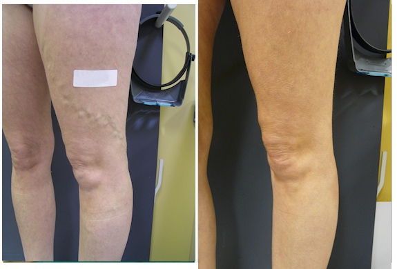
Case Study 1
Spider veins on the lateral thighs
This 41 year old lady presented with right sided lateral thigh spider veins which became more pronounced with the birth of her 4th child. She was more concerned with the unsightly appearance rather than having any significant leg symptoms. She wanted to get into shape and address her leg concerns after having completed her family. On examination we noted a cluster of spider veins in the upper thigh but there was the suspicion of a hidden feeding vessel which we located with a duplex ultrasound examination. We treated the leg by firstly closing down the feeding vessel with Ultrasound Guided Sclerotherapy (UGS) after which we were then able to treat the visible spider veins with Microsclerotherapy / Direct Vision Sclerotherapy (DVS). The post treatment photograph was taken 9 weeks after the initial course of 3 treatments which demonstrates an excellent outcome. It is possible that this patient may require in the future some small treatments with Microsclerotherapy / DVS to maintain this good outcome.

Case Study 2
Varicose Veins 1
This 38 year old presented with small varicose veins behind the left knee area. She was concerned regarding the unsightly appearance but was also starting to get some ache in the area. I had to firstly identify whether there was any underlying hidden veins and we proceeded with a duplex ultrasound examination. This revealed a small feeding vessel that would be amenable to Ultrasound Guided Sclerotherapy (UGS) and Endovenous Laser Treatment (ELT) was not required. We proceeded with Ultrasound Guided Sclerotherapy (UGS) and Modified Ambulatory Phlebectomy with only a single treatment being required. We did require a follow up treatment where some trapped blood was released to reduce the risk of possible pigmentation. The post treatment photograph was taken 6 weeks later demonstrating no significant visible scarring.

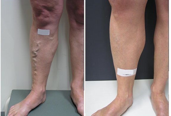
Varicose Veins 2
This 42 year old lady presented with a large bulging vein wrapping around the left outer thigh. She was concerned from both a symptomatic viewpoint as she was experiencing considerable throbbing pain and ache but was also concerned with the unsightly cosmetic appearance. She is a busy mother and could not afford any downtime. Duplex Ultrasound examination revealed a short and small feeding vein hiding beneath the skin that was directly responsible for the visible bulging vein in the outer thigh. The best treatment option for this presentation was a combination of Ultrasound Guided Sclerotherapy (UGS) and Modified Ambulatory Phlebectomy (MAP) performed in the one session. This procedure took 60 minutes to perform and the patient was able to immediately walk and return to normal duties with the provision she avoided heavy physical exercise for 7 days. The patient was informed that some temporary bruising was to be expected and some localised discomfort that usually responds to taking Nurofen if required. The post treatment photograph was taken 8 weeks after the treatment which demonstrates an excellent outcome. Although I do not expect to have to see this patient in the foreseeable future it is possible a small maintenance treatment will be required periodically should new veins develop.
Case Study 3
Massive varicose veins
This 42 year old male presented with a massive vein that extended from the upper inner thigh into the lower inner leg. A detailed history was taken which revealed that this grossly abnormal vein pattern was causing not only considerable leg ache, pain and throbbing but also resulted in frequent cramps and was quite frankly unsightly. Therefore the patient was concerned from both a symptomatic and cosmetic viewpoint. We proceeded to a Duplex Ultrasound Examination so that we can visualise any hidden abnormal veins and identify the of abnormal flow pattern. This revealed that there was a very large hidden abnormal vein that was directly responsible for the visible varicose veins on the inner thigh and calf. Discussion then occurred where it was advised the best treatment for this presentation was to offer a combination of treatment methods that are performed in the one session. These included EVLT (Endovenous Laser Ablation) to the large hidden vein which was then followed by UGS (Ultrasound Guided Sclerotherapy) to the visible veins which were then removed by MAP (Modified Ambulatory Phlebectomy). The post treatment photograph was taken 8 days after the treatment. This gentleman required two follow up visits to release some trapped blood which is performed under local anaesthetic for minimal discomfort. His leg will continue to improve over the next few weeks. As he has a significant genetic weakness to develop abnormal veins I expect that he will require some maintenance sclerotherapy (injection treatments) in the future as new veins may develop. Maintenance treatment can negate the requirement for the more involved and expensive EVLT and AMP treatments.

Case Study 4
Medium sized veins and spider veins on outer leg
This 59 year old lady presented with extensive reticular (medium sized blue veins) and spider veins on the outer right leg. She was concerned with the unsightly appearance but also had vague leg sensations of ache and stinging which were far beyond providing nuisance value. We proceeded to Duplex Ultrasound Examination which revealed no significant hidden veins but there were significant feeding vessels directly producing the appearance of the unsightly spider vein networks. Discussion followed that the best treatment for this presentation was Direct Vision Sclerotherapy (DVS). She was then informed that following treatment it was possible she may develop some small areas of pigmentation where there were large clusters of spider veins. These tend to fade completely within a few months. The patient proceeded with DVS and in all 3 treatment sessions were required for a successful outcome. The post treatment photograph was taken 3 months after the initial course of treatment was finished. Such vein networks usually mean the patient has a tendency to develop veins with time. The patient was informed that it was likely that periodic treatment would be required to maintain her excellent outcome.

Case Study 5
Varicose veins back of thigh
This 32 year old woman presented with large posterior thigh veins which developed after her 2nd pregnancy but became worse with her 3rd pregnancy. Having completed her family she was motivated to getting back to her pre pregnancy weight and fitness and wanted to address the unsightly veins which was starting to cause pain and throbbing. She was very frightened about having any surgical procedure and was wondering whether her leg veins could be treated with injection techniques. We needed to firstly perform a Duplex Ultrasound Examination which revealed the cause of her vein was coming from buttock veins that link into the pelvis and usually develop with pregnancy. This vein is amenable to Ultrasound Guided Sclerotherapy (UGS) and Microsclerotherapy provided we follow up with a few sessions of releasing trapped blood under local anaesthetic for minimal discomfort. This prevents the risk of developing prominent pigmentation that can occur in such presentations when treated with only injection techniques. Although this vein could also be treated with combination of Sclerotherapy and Modified Ambulatory Phlebectomy, which reduces the risk of pigmentation, the patient had a morbid fear of the minor surgical nature of the latter component. This presentation only required a single injection treatment and 3 sessions of releasing trapped blood. The post photograph was taken 10 weeks after the initial treatment. We were able to achieve the result the patient was hoping for and without any pigmentation. As always follow maintenance treatment may be required.

Case Study 6
Varicose Veins
This 54 year old woman presented with large single vein coursing down the front of the right leg. She did have symptoms of throbbing and ache but in particular hated the look of them as she was very active with many sporting interests and it was becoming increasingly embarrassing exposing her legs. We performed a venous duplex scan to evaluate the source of the pressure causing her visible varicose veins. On the right there was a very large hidden veins that in the past could only be treated with stripping surgery. The visible varicose vein which lies over the shin is particularly difficult to remove. The best treatment option for this presentation was to perform Endovenous Laser Ablation which was followed with Ultrasound Guided Sclerotherapy (UGS) to the vein in the calf which was then compressed with padding under the stocking. The patient required to have some trapped blood released under local anaesthetic for minimal discomfort. The after photograph was taken 4 months after initial single treatment. I recently reviewed the patient some 8 years after this treatment to treat some small spider veins. There was no evidence of any regrowth in this case.
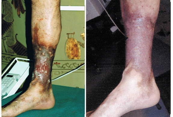
Case Study 7
Massive Leg Ulcer
This 61 year old man presented with a painful right leg which had significant skin changes and a large leg ulcer that reflects longstanding vein pressure. The black skin represents iron pigment that has been deposited into the skin due to blood leaking into the tissues. With time this breaks down the skin. A venous duplex scan revealed a massive hidden vein inside the leg directly passing through the skin changes. There were also some hidden veins near the ulcer that originate from deep within the muscle. The best treatment in my view was to provide Endovenous Laser Ablationto the hidden segments and then Ultrasound Guided Sclerotherapy (UGS) to residual hidden branches that could not be lasered. The after picture was taken 12 months after treatment which shows that the ulcer has healed and the skin was looking more nutritious and less pigmented. He is most likely going to require some top up treatment with injection techniques to prevent new veins from breaking down. Treating varicose veins early can prevent such ulcers from forming.